Fetal RhD and other red cell antigen genotyping using cell-free DNA (cfDNA) from maternal plasma provides a non-invasive means of predicting fetal red cell antigen status from antigen-defining genetic sequences. Its clinical role depends on the indication.
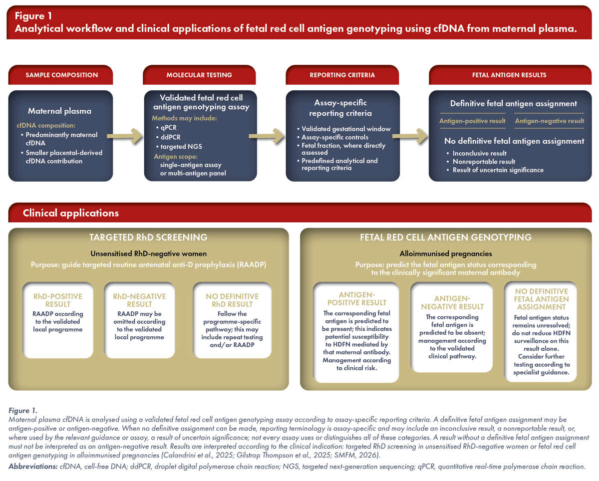
In unsensitised RhD-negative women, fetal RhD genotyping supports prevention of RhD sensitisation by guiding routine antenatal anti-D prophylaxis (RAADP). In pregnancies complicated by red cell alloimmunisation (hereafter, alloimmunised pregnancies), fetal red cell antigen genotyping determines whether the fetus carries the antigen corresponding to a clinically significant maternal antibody and therefore whether the fetus may be susceptible to haemolytic disease of the fetus and newborn (HDFN) mediated by that antibody. These applications require different laboratory pathways: high-throughput fetal RhD genotyping for targeted RAADP is designed for population screening, whereas fetal red cell antigen genotyping in alloimmunised pregnancies is performed within specialist diagnostic services (Figure 1). Accordingly, assay selection, the validated gestational window, reporting criteria and downstream clinical management vary with the clinical indication and available testing platform (Regan et al., 2025; Moise et al., 2025; SMFM, 2026).
Biological Basis and Sample Adequacy
Maternal plasma contains predominantly maternal cfDNA together with a smaller cfDNA fraction released mainly by placental trophoblasts, which normally reflects the fetal genotype at inherited blood group loci. Fetal red cell antigen genotyping therefore analyses cfDNA from maternal plasma rather than fetal blood directly (Yuen et al., 2024).
The assay detects sequence information in maternal plasma cfDNA and infers fetal genotype from the placental contribution to the sample; it does not directly measure antigen expression on fetal red blood cells. The reported fetal antigen status is therefore inferred from the targeted genotype and may be affected by variant alleles that alter the relationship between genotype and serological phenotype (Yuen et al., 2024; Dufour et al., 2022; SMFM, 2026).
Fetal fraction, which generally refers to the proportion of maternal plasma cfDNA attributable to the placental contribution, is particularly relevant when an antigen-negative result is inferred from absence of the target sequence. Such a result is reliable only when testing is performed within the assay’s validated gestational window and its sample-adequacy and reporting criteria are met. Fetal fraction and sample adequacy are related but not interchangeable: some assays estimate fetal fraction quantitatively, whereas others establish reportability using fetal-specific markers, internal controls or other assay-specific criteria (Calandrini et al., 2025; Gilstrop Thompson et al., 2025; SMFM, 2026).
Approaches to establishing sample adequacy include Y-chromosome markers in pregnancies with a male fetus, methylated RASSF1A as a sex-independent placental marker and, in targeted sequencing assays, informative single-nucleotide or insertion/deletion polymorphisms. In a study evaluating methylated RASSF1A as a sex-independent control, the marker helped identify samples in which an absent RHD signal could reflect insufficient placental contribution to maternal plasma cfDNA; in such samples, the absent signal should not be interpreted as a definitive fetal RhD-negative result (Chan et al., 2006). Methylated RASSF1A therefore functions as a sample-adequacy control rather than as an antigen-defining target.
Gestational age and pre-analytical handling also influence sample adequacy and test performance. The placental cfDNA contribution generally increases with gestation, which is why each assay requires a validated earliest testing window. Sample processing is equally relevant: cellular contamination and differences in plasma-processing protocols can increase the maternal DNA background and alter the relative placental cfDNA contribution, so standardised blood-processing and plasma-separation procedures form part of test quality (Chiu et al., 2001).
Molecular Methods
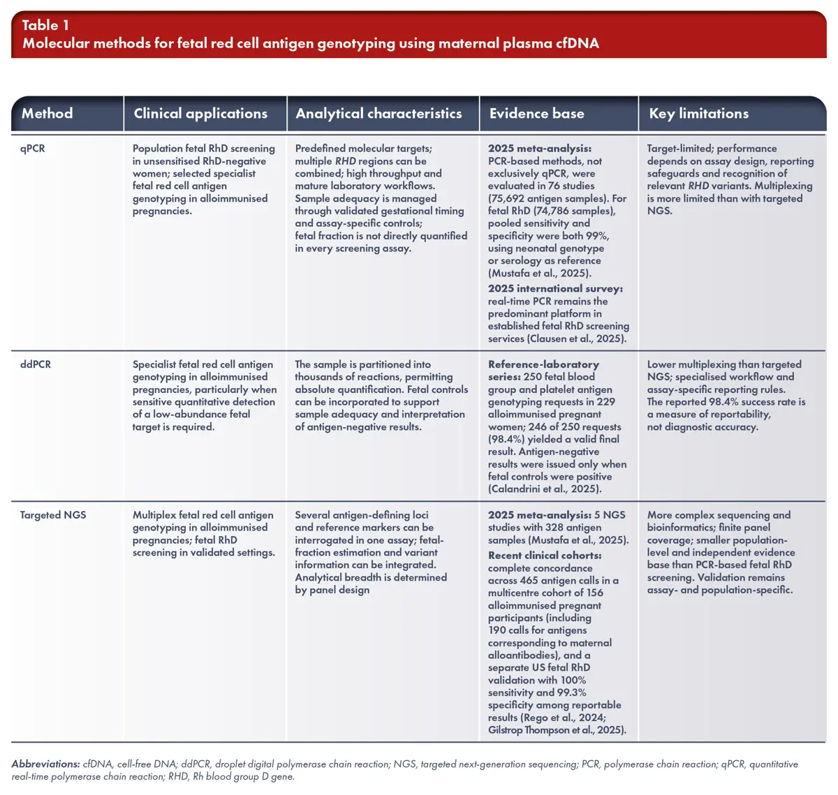
Quantitative real-time polymerase chain reaction (qPCR), droplet digital polymerase chain reaction (ddPCR) and targeted next-generation sequencing (NGS) are all used for fetal red cell antigen genotyping. These platforms serve complementary rather than sequential roles. They differ in sample throughput, the number and type of antigen-defining loci that can be interrogated, sensitivity for low-abundance alleles, capacity to incorporate fetal-fraction and variant information, laboratory complexity, and the extent of clinical validation (Table 1).
qPCR has the largest programme-level evidence base for fetal RhD screening. High-throughput assays usually interrogate more than one RHD region and are designed to process large numbers of samples. Their main advantages are extensive validation, scalability and long-standing use in population-screening programmes. Interpretation remains limited to the targets included in the assay, so performance depends on assay design and recognition of relevant RHD variants.
ddPCR partitions a sample into thousands of individual reactions and permits sensitive, absolute quantification of low-abundance targets. It is particularly useful in specialist antigen-specific testing, where a fetal allele must be detected against a large maternal background. Fetal controls can be incorporated to support sample adequacy and interpretation of antigen-negative results; assay performance and reporting rules remain laboratory-specific (Calandrini et al., 2025).
Targeted NGS permits simultaneous interrogation of several antigen-defining loci and multiple reference markers within one assay. The number of loci is determined by panel design rather than by a fixed limit of the technology. This makes NGS particularly useful when several clinically relevant antigens or complex RHD patterns must be assessed and when fetal-fraction estimation is integrated into the same workflow. Its evidence base is expanding but remains much smaller than that for polymerase chain reaction (PCR)-based fetal RhD genotyping. Recent United States (US) and European studies have reported high concordance for reportable NGS results, but assay-specific validation, population representation and independent evidence remain important considerations (Rego et al., 2024; Gilstrop Thompson et al., 2025; Emahazion et al., 2025; SMFM, 2026).
Evaluation of fetal red cell antigen genotyping requires distinction between analytical validity, clinical validity and clinical utility. Analytical validity concerns accurate and reproducible detection of the intended molecular target, whereas clinical validity concerns agreement between the reported fetal antigen result and established fetal or neonatal antigen status. Clinical utility concerns whether use of the test changes management in a way that improves care in the healthcare setting concerned. High analytical or clinical validity does not by itself establish clinical utility across healthcare systems (Mustafa et al., 2025; Regan et al., 2025; SMFM, 2026).
Clinical Applications and Timing
Within validated screening programmes, fetal RhD genotyping guides targeted RAADP in unsensitised RhD-negative women by establishing fetal RhD status and thereby whether RAADP is indicated within that programme. In alloimmunised pregnancies, the maternal antibody determines the fetal antigen target. A definitive antigen-negative result supports absence of the corresponding fetal antigen and therefore indicates that HDFN mediated by that antibody is not expected; it does not exclude risk associated with other clinically significant maternal antibodies or non-immunological causes of fetal anaemia. A definitive antigen-positive result indicates potential susceptibility to HDFN mediated by that antibody but does not by itself diagnose fetal anaemia or HDFN. Subsequent surveillance and further testing depend on antibody specificity, concentration or titre, obstetric history and specialist assessment (Regan et al., 2025; Moise et al., 2025).
Timing is assay-, indication- and programme-specific. International implementation of high-throughput fetal RhD screening is not confined to a single gestational window. In a 2025 international survey led by the ISBT cfDNA subgroup, 52 laboratories from 22 countries responded; 38 performed fetal RhD screening and 27 of these 38 reported routine nationwide programmes. Testing ranged from 8 to 27 weeks, most commonly around 10–12 or 24–27 weeks (Clausen et al., 2025). BSH considers high-throughput fetal RhD screening sufficiently accurate from 11 weeks, while NICE reports poorer performance at or before 11 weeks. The Dutch national programme used centralised qPCR testing at 27 weeks and reported 99.94% sensitivity with a 0.03% false-negative rate (de Haas et al., 2016). A recent US clinical validation study of an NGS assay reported 100% sensitivity and 99.3% specificity among reportable results from 9 weeks or later (Gilstrop Thompson et al., 2025). These figures are not directly comparable because the studies differed in assay, gestational timing, population, comparator and handling of nonreportable results. The reported windows therefore reflect assay validation and programme design rather than a single universal optimum (Regan et al., 2025; NICE, 2016).
In alloimmunised pregnancies, timing is antigen- and guidance-specific. BSH specialist guidance uses reference-laboratory-specific windows: RHD and RHCE testing are performed at the gestation advised by the reference laboratory, with supporting data for RHD and RHCE (c) testing at or after 16 weeks; KEL*01 testing should not be performed before 20 weeks and repeat testing after an apparently K-negative result may be advised. By contrast, a 2025 US multidisciplinary guideline recommends cfDNA testing after 10 weeks for alloimmunised pregnancies with anti-D, anti-C, anti-c, anti-E, anti-K or anti-Fyᵃ. SMFM 2026 is more conservative: when the reproductive partner or sperm donor is heterozygous for the relevant antigen or unavailable for testing, amniocentesis remains the standard of care for fetal genotype evaluation; cfDNA may be offered after counselling if diagnostic testing or empiric HDFN monitoring is declined. Accordingly, no single gestational timing rule applies across these different clinical and guidance frameworks. (Table 2; Regan et al., 2025; Moise et al., 2025; SMFM, 2026).
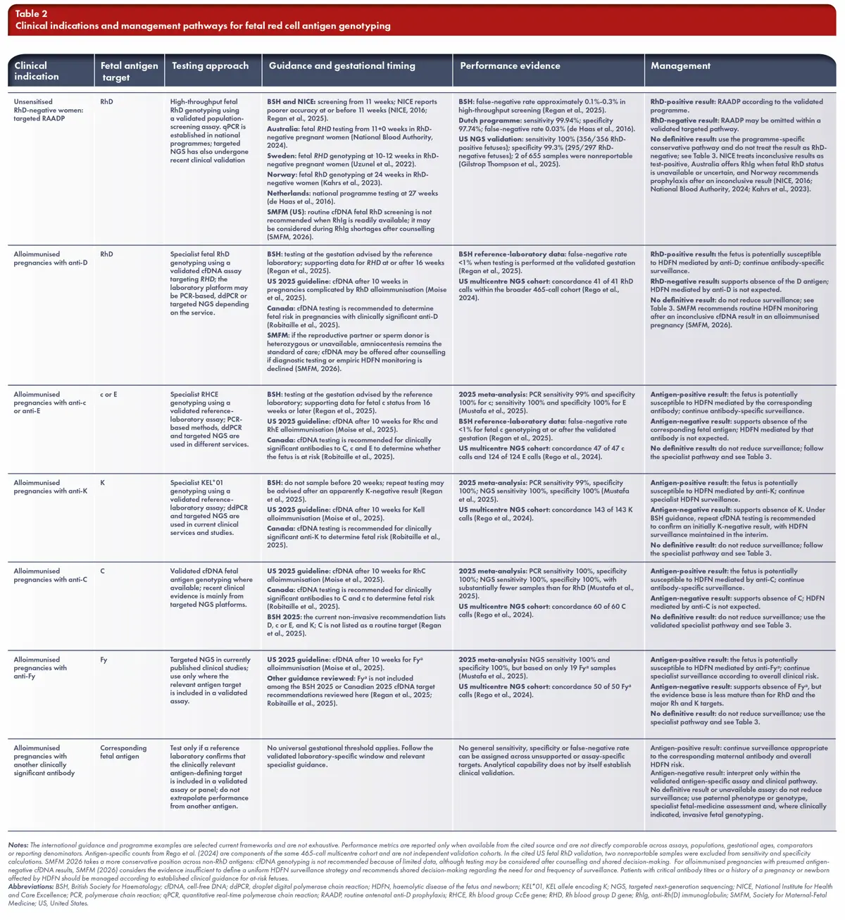
Evidence for fetal red cell antigen genotyping beyond RhD remains antigen- and assay-specific. Recent cohorts and the 2025 meta-analysis show high diagnostic accuracy for several targets, but the volume and maturity of evidence are not equivalent across antigens. Validation datasets for C, c, E and K are substantially smaller than those for RhD, while evidence for Fyᵃ remains particularly limited (Table 2). Inclusion of an antigen in a multi-antigen panel demonstrates analytical capability but does not by itself establish equivalent clinical validation, routine availability or guideline-supported use. Panel coverage and the validated clinical pathway must therefore be confirmed for the antigen corresponding to the maternal antibody before testing (Mustafa et al., 2025; Rego et al., 2024; Emahazion et al., 2025; Moise et al., 2025; SMFM, 2026).
Result Interpretation and Discordance
Terminology for results that do not provide a definitive fetal antigen assignment is not standardised across assays, laboratories or guidance. Published sources use overlapping terms for different analytical circumstances, and no cross-platform universal definitions exist. Table 3 summarises the terminology currently used across assays and guidance. The laboratory-reported term, the stated reason and the associated management guidance should be preserved. Irrespective of terminology, a result without a definitive fetal antigen assignment must not be interpreted as an antigen-negative result (Calandrini et al., 2025; Gilstrop Thompson et al., 2025; SMFM, 2026).
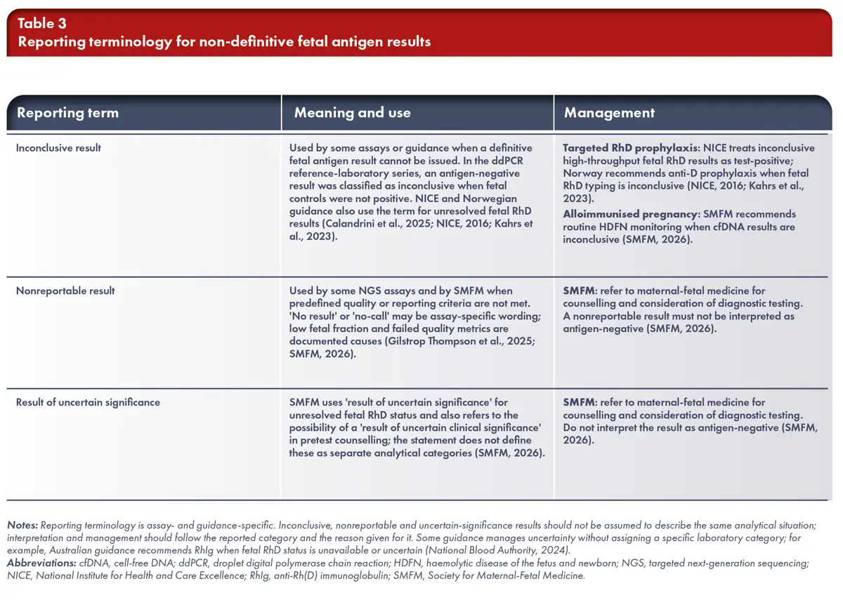
The clinical consequences of error are asymmetric. In a targeted RAADP pathway, a false-positive fetal RhD result generally leads to unnecessary RhIg, whereas a false-negative fetal RhD result can lead to omission of RAADP in a woman carrying an RhD-positive fetus. In alloimmunised pregnancies, erroneous antigen-negative results may inappropriately reduce HDFN surveillance. Reporting criteria and the management of results without a definitive fetal antigen assignment are therefore part of clinical safety, not merely technical detail (NICE, 2016; Regan et al., 2025; SMFM, 2026).
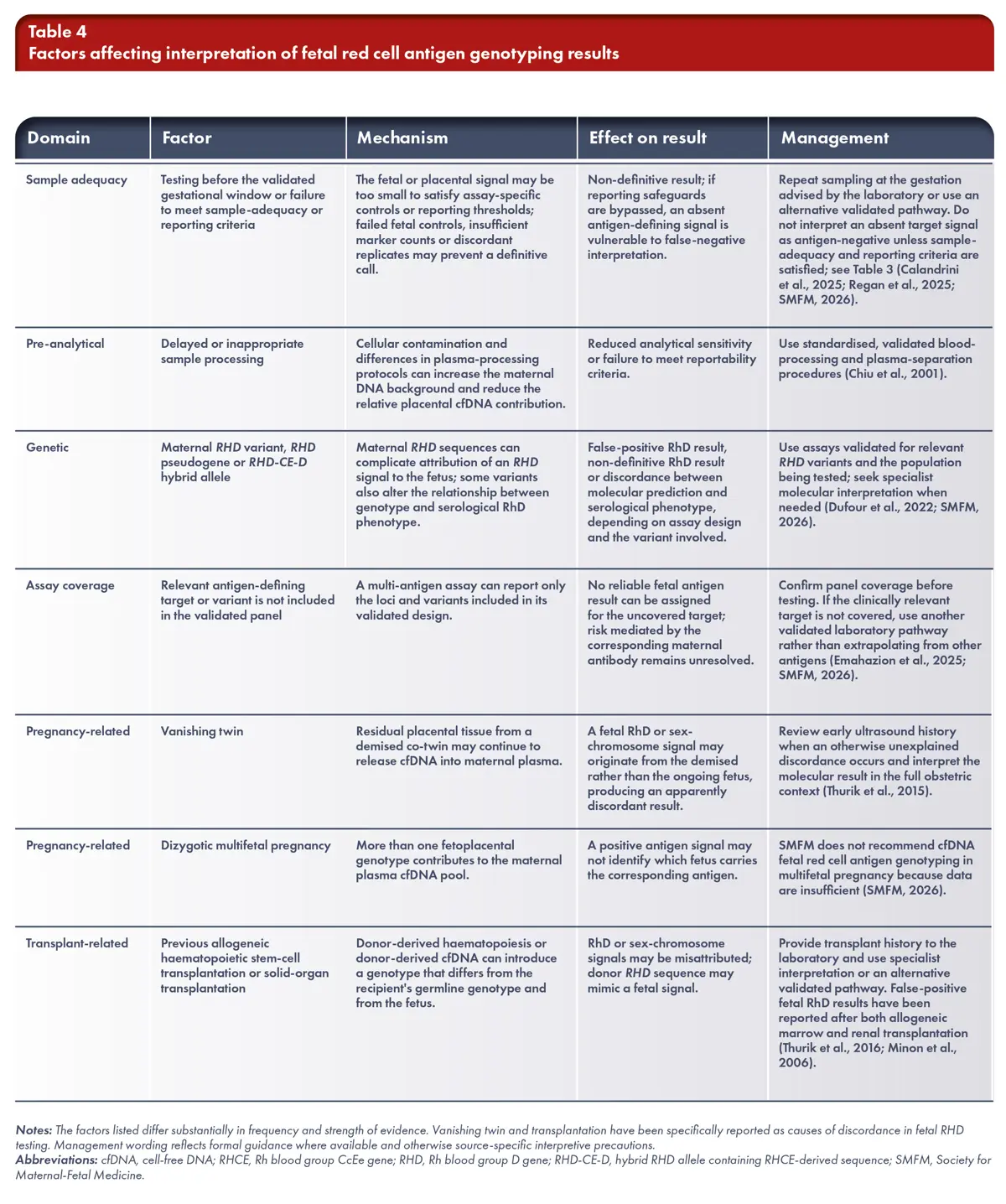
Discordant or non-definitive results can arise from assay or sample-adequacy limitations, pre-analytical handling, genetic or variant-related interference, incomplete panel coverage, pregnancy-related biological factors, or donor-derived DNA after transplantation (Table 4). These domains are used here as pragmatic groupings for interpretation rather than as universal laboratory categories. Population-specific RHD variation is particularly important because maternal RHD sequences can complicate attribution of an RHD signal to the fetus, and the relationship between an RHD sequence and serological RhD phenotype may vary with the underlying variant spectrum. Assay design and validation should therefore reflect the genetic diversity of the population in which testing is used; performance established in one population should not be transferred automatically to another (Dufour et al., 2022; SMFM, 2026).
International Implementation and Guidance
Clinical implementation differs across healthcare systems. International survey data show that fetal RhD screening is integrated into routine care in multiple countries, with European programmes providing the longest population-level experience; however, implementation data remain weighted towards Europe and high-income settings (Clausen et al., 2025). National programmes and clinical validation studies differ in assay design, gestational timing, reporting rules and healthcare context, so their performance estimates should not be treated as directly interchangeable. In the United States, recent NGS validation has shown high analytical performance from early gestation, whereas SMFM does not recommend routine cfDNA fetal RhD screening in unsensitised RhD-negative women when RhIg is readily available. These differences reflect programme design, available alternative strategies, cost-effectiveness, reimbursement and health-system policy as well as analytical performance (Gilstrop Thompson et al., 2025; SMFM, 2026).
Before using a cfDNA result to omit RAADP or modify HDFN surveillance, clinicians should confirm the specific laboratory service, antigen and variant coverage, validated gestational window, reporting criteria and management pathway for results without a definitive fetal antigen assignment. Postnatal confirmation remains valuable in specialist diagnostic pathways because comparison with neonatal blood group results can identify rare assay failures or previously unrecognised variants and strengthen real-world quality assurance (Regan et al., 2025). Clinical implementation should also be supported by documented laboratory quality processes, including validated pre-analytical procedures, internal controls, defined policies for nonreportable results and, where available, participation in external quality assessment (Chiu et al., 2001; Clausen et al., 2025; Regan et al., 2025).
Conclusions
Fetal RhD genotyping using maternal plasma cfDNA is an established non-invasive strategy in validated population-screening and specialist diagnostic pathways. Its clinical value lies not simply in detecting an RHD sequence, but in matching the clinical indication and fetal antigen target to an assay that is validated for the relevant gestational window, population and reporting framework. Genotyping of other clinically significant fetal red cell antigens is increasingly feasible, but the evidence base and degree of implementation remain less mature and more assay-specific than for RhD.
qPCR has the broadest population-level evidence for fetal RhD screening; ddPCR supports sensitive quantitative testing in specialist settings; and targeted NGS enables broader multi-antigen interrogation with multiple reference markers and, in some assays, integrated fetal-fraction assessment. These approaches are complementary rather than sequential, and no platform is universally preferable. Analytical breadth should therefore not be equated with equivalent clinical validation across antigens or healthcare settings.
For unsensitised RhD-negative women, fetal RhD genotyping can support targeted RAADP where this strategy is validated and implemented. In alloimmunised pregnancies, fetal red cell antigen genotyping can refine HDFN surveillance by establishing whether the fetus is predicted to carry the antigen corresponding to the maternal antibody. Across both settings, clinical safety depends on satisfying assay-specific sample-adequacy and reporting criteria, recognising relevant variants and having an explicit pathway for results without a definitive fetal antigen assignment, which must not be interpreted as antigen-negative. As testing expands beyond RhD, analytical validity, clinical validity, quality assurance and guideline-supported use should remain clearly distinguished.
References
- Regan F, Veale K, Robinson F, Brennand J, Massey E, Qureshi H, et al. Guideline for the investigation and management of red cell antibodies in pregnancy: A British Society for Haematology guideline. Transfus Med. 2025;35(1):3-23. doi:10.1111/tme.13098. Correction in: Transfus Med. 2025;35(6):593. doi:10.1111/tme.70013.
- National Institute for Health and Care Excellence. High-throughput non-invasive prenatal testing for fetal RHD genotype. HealthTech guidance HTG420. London: NICE; 2016. Last reviewed 2021.
- Yuen N, Lemaire M, Wilson SL. Cell-free placental DNA: What do we really know? PLoS Genet. 2024;20(12):e1011484. doi:10.1371/journal.pgen.1011484.
- Chan KCA, Ding C, Gerovassili A, Yeung SW, Chiu RWK, Leung TN, et al. Hypermethylated RASSF1A in maternal plasma: A universal fetal DNA marker that improves the reliability of noninvasive prenatal diagnosis. Clin Chem. 2006;52(12):2211-2218. doi:10.1373/clinchem.2006.074997.
- Chiu RWK, Poon LLM, Lau TK, Leung TN, Wong EMC, Lo YMD. Effects of blood-processing protocols on fetal and total DNA quantification in maternal plasma. Clin Chem. 2001;47(9):1607-1613.
- Clausen FB, Hellberg A, Toly-Ndour C, Nielsen ET, de Haas M; Noninvasive Fetal Antigen Genotyping Survey Group. Noninvasive fetal antigen genotyping: Results from a survey on the status of clinical implementation. Vox Sang. 2025;120(9):935-943. doi:10.1111/vox.70062.
- Mustafa HJ, Najjariasl P, Aghajani F, Sambatur EV, Khalil A, Moise KJ Jr, et al. Diagnostic accuracy of cell-free DNA for the determination of fetal red blood cell antigen genotype: a systematic review and meta-analysis. Am J Obstet Gynecol. 2025;233(5):428-445.e16. doi:10.1016/j.ajog.2025.05.004.
- Calandrini C, Verhagen OJHM, Tissoudali A, Homburg CHE, Vessies J, Brussee M, et al. Real-world performance of a clinical droplet digital polymerase chain reaction assay for non-invasive foetal blood group and platelet antigen genotyping of alloimmunized pregnant women with antibodies directed against RhD, RhE, Rhc, RhC, K1, HPA-1a or HPA-5b: A 1-year experience. Vox Sang. 2025;120(2):170-177. doi:10.1111/vox.13777.
- Rego S, Ashimi Balogun O, Emanuel K, Overcash R, Gonzalez JM, Denomme GA, et al. Cell-Free DNA Analysis for the Determination of Fetal Red Blood Cell Antigen Genotype in Individuals With Alloimmunized Pregnancies. Obstet Gynecol. 2024;144(4):436-443. doi:10.1097/AOG.0000000000005692.
- Gilstrop Thompson M, Xu W, Moore B, Wang T, Sun N, Pewar H, et al. Clinical Validation of a Prenatal Cell-Free DNA Screening Test for Fetal RHD in a Large U.S. Cohort. Obstet Gynecol. 2025;145(2):211-216. doi:10.1097/AOG.0000000000005794.
- Emahazion T, Badri A, Jalkesten E, Flodstrom M, Ajne G, Uzunel M, et al. Next-generation sequencing-based approach for fetal DNA quantification and blood antigen detection in alloimmunized pregnancies. Am J Obstet Gynecol MFM. 2025;7(7):101694. doi:10.1016/j.ajogmf.2025.101694.
- Moise KJ Jr, Markham KB, Spinella PC, Sherwood MR, Robinson KA, Wilson LM, et al. A Clinical Practice Guideline for the Management of Pregnancy Alloimmunized to Red Blood Cell Antigens. JAMA Netw Open. 2025;8(11):e2544649. doi:10.1001/jamanetworkopen.2025.44649.
- Society for Maternal-Fetal Medicine (SMFM), Rink BD, Kuller JA, Sparks TN, Prabhu M, Mardy AH, et al. Society for Maternal-Fetal Medicine Statement: Evaluation and management of cell-free DNA screening for fetal red cell antigen genotype in alloimmunized and non-alloimmunized pregnancies. Pregnancy (Hoboken). 2026;2(4):e70339. doi:10.1002/pmf2.70339.
- Dufour P, Gerard C, Chantraine F, Minon JM. Investigation of discrepancies obtained during 15 years of non-invasive fetal RHD genotyping in apparent serologic RhD-negative pregnant women. Prenat Diagn. 2022;42(10):1262-1272. doi:10.1002/pd.6219.
- Thurik FF, Ait Soussan A, Bossers B, Woortmeijer H, Veldhuisen B, Page-Christiaens GCML, et al. Analysis of false-positive results of fetal RHD typing in a national screening program reveals vanishing twins as potential cause for discrepancy. Prenat Diagn. 2015;35(8):754-760. doi:10.1002/pd.4600.
- Thurik FF, Page-Christiaens GCML, Ait Soussan A, Ligthart PC, Cheroutre GMAF, Bossers B, et al. Fetal RHD genotyping after bone marrow transplantation. Transfusion. 2016;56(8):2122-2126. doi:10.1111/trf.13669.
- Minon JM, Senterre JM, Schaaps JP, Foidart JM. An unusual false-positive fetal RHD typing result using DNA derived from maternal plasma from a solid organ transplant recipient. Transfusion. 2006;46(8):1454-1455. doi:10.1111/j.1537-2995.2006.00916.x.
- de Haas M, Thurik FF, van der Ploeg CPB, Veldhuisen B, Hirschberg H, Ait Soussan A, et al. Sensitivity of fetal RHD screening for safe guidance of targeted anti-D immunoglobulin prophylaxis: prospective cohort study of a nationwide programme in the Netherlands. BMJ. 2016;355:i5789. doi:10.1136/bmj.i5789.
- National Blood Authority. Guideline for the prophylactic use of Rh D immunoglobulin in pregnancy care. Canberra: National Blood Authority; 2024.
- Uzunel M, Tiblad E, Mörtberg A, Wikman A. Single-exon approach to non-invasive fetal RHD screening in early pregnancy: An update after 10 years’ experience. Vox Sang. 2022;117(11):1296-1301. doi:10.1111/vox.13348.
- Kahrs BH, Tiller H, Haugen G, Bakke K, Akkök ÇA. Alloimmunisering mot erytrocytt-antigener. Veileder i fødselshjelp. Norsk gynekologisk forening; revised April 2023.
- Robitaille N, Lieberman L, Clarke G, Barrett J, De Vrijer B, Hume HA, et al. National Consensus Statements for the Prevention of Maternal Rhesus D Alloimmunization and Management of Alloimmunized Pregnancies: A Modified Delphi Process. J Obstet Gynaecol Can. 2025;47(10):103113. doi:10.1016/j.jogc.2025.103113.